Figures & data
Table 1 The 2015 International Panel for NMOSD Diagnostic Criteria for Adult Patients
Figure 1 T2-weighted mid-sagittal image centered at T3. Long contiguous segment (T3 to T6) central T2 hyperintense lesion mildly expanding the cord.
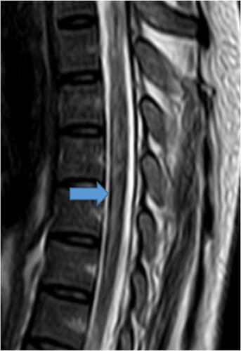
Figure 2 T2-weighted mid-sagittal image proximal to the level on previous image shows long segment expansion of the central canal (syrinx).
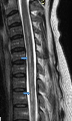
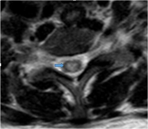
Figure 3 Axial T2-weighted image at the level of T3 revealed central cord hyperintensity.