
Figures & data
Figure 1 Physical examination showing eunuchoid body proportions, obesity, long extremities, gynecomastia, scanty pubic hairs, small testes (A and B). Shallow ulcer with yellow necrotic tissue on the lower-left leg and atrophie blanche on both lower legs (C). Varicose veins with hyperpigmented indurated skin appear on both lower legs (D and E).
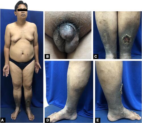
Figure 2 Histopathological findings (Hematoxylin and eosin; x100 and x200 magnifications) showed epidermal acanthosis, dermal fibrosis, and thickening, hemosiderin deposits (blue arrow).
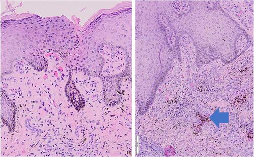
Figure 3 Fibrin clot at the center of vacutainer (red arrow) (A). Four PRF clots were placed in the entire ulcer area (B). Clinical manifestation of venous leg ulcer before (C), after 4 weeks (D), and after 7 weeks of PRF dressing (E).
