Figures & data
Figure 1 (A) Volumetric-rendering reconstructed image of the chest CT shows the path of the epidural catheter (arrows labeled #1) crossing the midline between the T6-7 spinous processes before entering the pleural space via the left 6th-7th intercostal space. (B) Maximal intensity projection (MIP) coronal CT image shows the course of the catheter (arrows labeled #2) enters the left 6th-7th intercostal space. (C) MIP axial CT image shows the course of the catheter (arrow labeled #3), crossing the midline and entering the pleural space. Note the tip of the catheter immediately medial the chest tube (arrowhead labeled #4). (D) MIP sagittal CT image demonstrates the catheter (arrow labeled #5) anchored at the site of entering the pleural space.
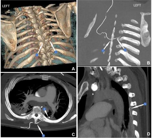
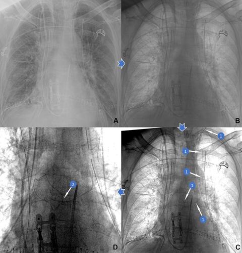
Figure 2 Series of chest x-ray digital image enhancements reveal intrapleural epidural misplacement in our patient. These chest films were obtained on day after epidural placement, 3 days prior to the CT in . (A) Initial chest radiograph. (B) The same image with grayscale inversion. (C) After adjusting image window and level (digital contrast enhancement), the path of the epidural catheter is better seen (arrows labeled #1). (D) A magnified crop of the radiograph highlights the path of the epidural catheter crossing midline with tip in the left lung field (arrow labeled #2) abutting against the left chest tube (arrows labeled #3).
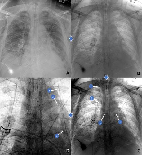
Figure 3 Series of chest x-ray digital image enhancements to reveal the appropriate TEP in another bilateral orthotopic lung transplant patient. (A) The initial chest radiograph taken within a day after the epidural placement. (B) The same image with grayscale inversion. (C) After adjusting image window and level (digital contrast enhancement), the path of the epidural catheter is better seen (arrows labeled #1). (D) A magnified crop of the radiograph highlights the path of the epidural catheter with tip eventually overlying the midline vertebral body (arrow labeled #2).