Figures & data
Table 1 Summary of Pertinent Investigation Results of the Case
Figure 1 Colonoscopy findings of a-12-year-old male adolescent from Ethiopia: Diffuse colon mucosal inflammation with intense rectal inflammation with extensive ulceration and exudates and loss of vascular patterns, and biopsied with the impression of Ulcerative colitis rule out IBD.
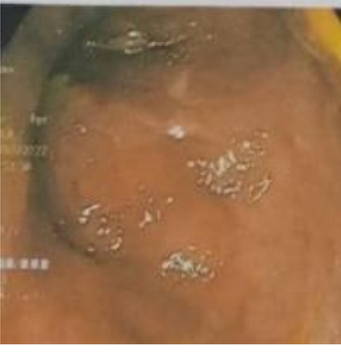
Figure 2 Low power photomicrograph revealing fragments of mucosal biopsy with foci of lymphoplasmacytic (yellow arrow) lamina propria infiltration, hemorrhage (red arrows) and a focus of an egg of S. mansoni (down blue arrow).
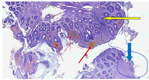
Figure 3 High power photomicrograph showing eggs of S.mansoni (down arrows) with surrounding inflammatory cells.
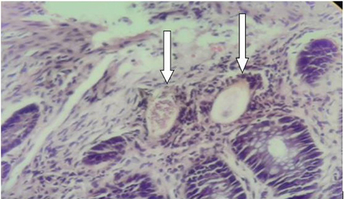
Figure 4 High power photomicrograph showing an egg of S. mansoni (down arrow) surrounded by mixed inflammatory cells and multinucleated giant cell (red arrow) with adjacent fibrosis.
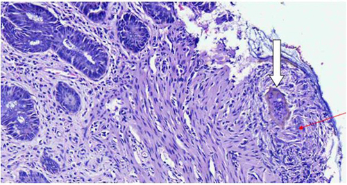
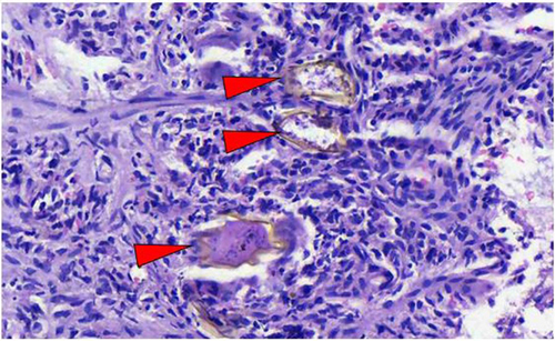
Figure 5 High power photomicrograph revealing eggs of S. mansoni (red arrow heads) surrounded by mononuclear inflammatory cells in the lamina propria.