Figures & data
Figure 1 Painless single ulcer, with a red base, white edges on the palate (A). Lesions healed in second visit, 14 days later (B).
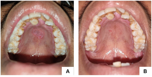
Figure 2 White plaque with erythematous area on the palate, buccal mucosa right and left, and tongue (A–E). Fissured on the corners of the lip. Second visit (three weeks), the lesions disappeared (F–J).
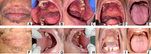
Figure 3 Greyish white plaques on mandibular anterior gingiva, erosion on lower labial mucosa (A). Erosion on left buccal mucosa (B). Erosion on left buccal mucosa (C). The lesion has improved (7 days) (D–F).
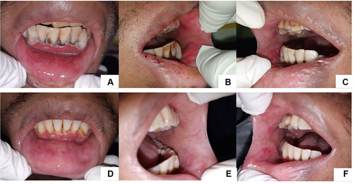
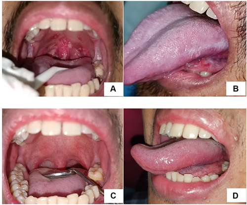
Figure 4 Multiple ulcers on soft palate, oropharynx, uvula (A), single ulcer on ventrolateral of the tongue and white corrugated, adherent plaques on left lateral of the tongue (B) two months after first visit, the lesions improved (C and D).