Figures & data
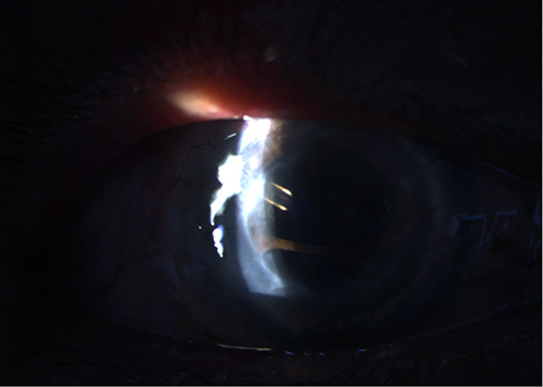
Figure 1 Failed graft after repeat penetrating keratoplasties. The patient complained of a significant decrease in vision, redness, grittiness, and pain.
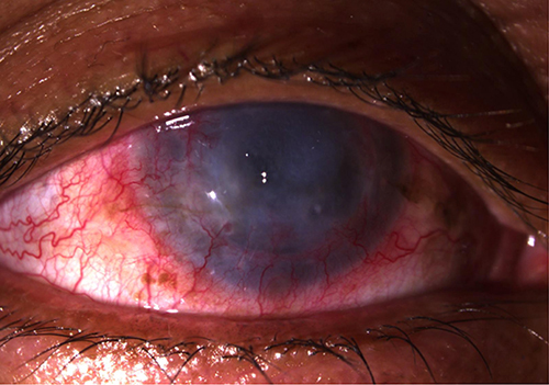
Figure 2 Patient one-week post-trabeculectomy with clear graft.
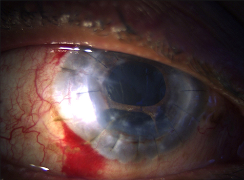
Figure 3 Thin, avascular bleb and no leak at one-month follow-up post-trabeculectomy with mitomycin C.
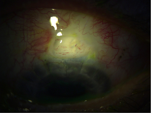
Figure 4 Scleral thinning 22-months post-trabeculectomy with mitomycin C.
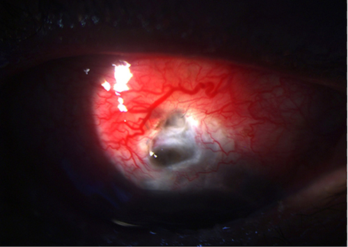
Figure 5 Bleb leak with positive Seidel test viewed after fluorescein staining.
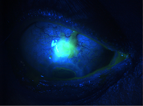
Figure 6 One-week post bleb revision with scleral patch.
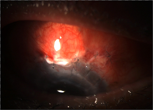
Figure 7 Clear graft six months after Ahmed tube implant positioned in the posterior chamber.
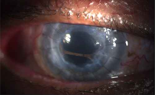
Figure 8 Tube positioned behind the iris with extensive peripheral anterior synechia.