Figures & data
Table 1 Cases 1–6 Including Age, Sex, Days Under Care, Comorbidities
Table 2 Pathogenesis of Disease Burden and Prevention Taken Respectively
Figure 1 Grade 3, concentric, corkscrew-type stenosis seen on CT chest (A) and pre-dilation bronchoscopy (B). Post-dilation bronchoscopy reveals airway patency (C).
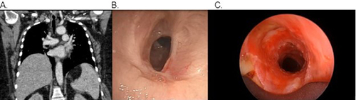
Figure 2 Grade 3, complex corkscrew-type stenosis which extended 3 tracheal rings seen on CT chest (A) and pre-dilation bronchoscopy (B). Post-dilation bronchoscopy reveals airway patency (C).
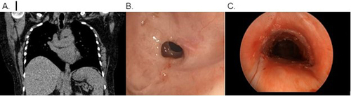
Figure 3 Grade 3, concentric, corkscrew-type stenosis which extended at least 2 tracheal rings seen on CT chest (A) and pre-dilation bronchoscopy (B). Post-dilation bronchoscopy reveals airway patency (C).
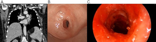
Figure 4 Grade 3, complex, concentric stenosis seen on CT chest (A) and pre-dilation bronchoscopy (B). Post-procedure bronchoscopy reveals airway patency with silicone studded hourglass stent in place (C).
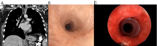
Figure 5 Grade 3, oval, corkscrew-type stenosis which extended 2 tracheal rings seen on CT chest (A) and pre-dilation bronchoscopy (B). Post-dilation bronchoscopy reveals airway patency (C).
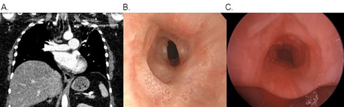
Figure 6 Grade 3, concentric stenosis seen on CT chest (A) and pre-dilation bronchoscopy (B). Post dilation bronchoscopy reveals airway patency (C).
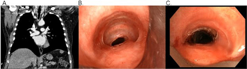
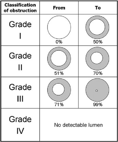
Figure 7 Classification grading demonstrating various levels of stenosis of the lumen based on the Meyer’s Cotton system.Citation15