
Figures & data
Table 1 Patient’s Characteristics
Figure 1 Thorax CT before treatment initiation. Intravenous contrast was administered prior to the exam. Extra pulmonary mass that infiltrates the thoracic cage and the ribs, as shown with the red arrow.
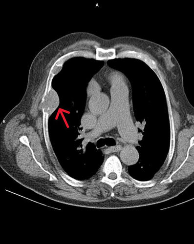
Figure 2 Biopsy from one of the osteolytic rib lesions shows bland fibroblastic spindle cell proliferation admixed with numerous osteoclastic giant cells(arrows), findings which in context are most suggestive of bone changes due to hyperparathyroidism (hematoxylin-eosin, x200).
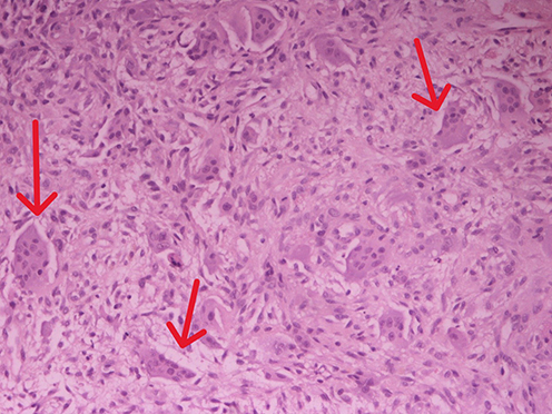
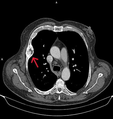
Figure 3 Thorax CT after i.v contrast, 18 months post treatment showed regression of the tumor in the thoracic cage (from 4.7×3.9 to 4.3×2.8 cm) and the ribs, as shown with the red arrow.
Box 1 Differential Diagnosis of Bone Lesions