Figures & data
Figure 1 Coronal and axial neck and chest CT scan shows asymmetric nodular thickening of the upper thoracic trachea (blue arrow) with a luminal narrowing >50% (A and B). Axial neck and chest CT scan shows asymmetric soft-tissue thickening of the intrathoracic trachea (yellow arrow) with significant airway narrowing (C).
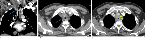
Figure 2 Vocal cord (green arrow) (A). Suprastomal granulation tissue (blue arrow) (B and C). Friable mass with ragged mucosa over the anterior and anterolateral part of the trachea around supracarinal area occluding ~60% of the lumen (yellow arrow) (D).
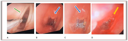
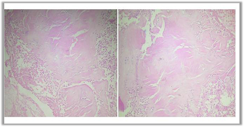
Figure 3 Section of tracheal sample showing acanthotic squamous epithelium with subepithelial deposition of pink hyaline extracellular material. Congo red stain was positive.