Figures & data
Figure 1 Erect abdomino pelvic x-ray (AP & lateral) showing radio opaque T-shaped material and intrauterine probe in the pelvis.
Notes: The white arrows indicate the T-shaped CU-IUD and the black arrows indicate the intrauterine probe.
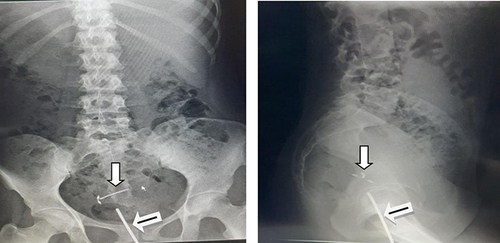
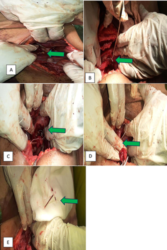
Figure 2 The removal process of CU-IUD from the left adnexa and uterine wall captured in pictures.
Notes: The green arrows indicate the details of the IUCD in the peritoneal cavity, specifically; (A) One of the CU-IUD’s arm is visible up on peritoneal entry, (B) CU-IUD string under the Fallopian tube: (C) Arrow pointing on the uterine fundus: (D) CU-IUD stem encased in the fallopian tube fimbriae: (E) Fully retrieved CU-IUD.