Figures & data
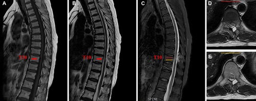
Figure 1 Magnetic resonance images (MRI) of pre-operation. (A–C) Preoperative sagittal MRI showed a sizeable IDEM tumor (red arrow) at the T10 level with an intact envelope and clear borders. (D and E) Preoperative axial MRI showed that the IDEM tumor significantly compressed the thoracic spinal cord to the right side, with 80% intraspinal encroachment. The red and yellow lines represent the axial planes in (C).
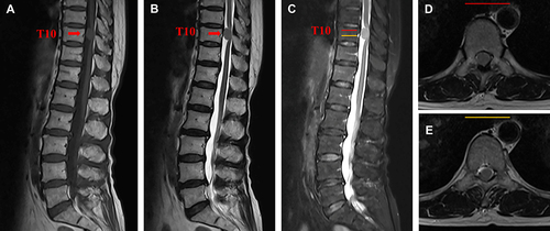
Figure 2 Intra-operative endoscopic images of UBE procedure. (A) Bleeding control and soft tissue detaching were performed with a bipolar radiofrequency instrument. (B) Intraoperative localization was performed using a 1.5mm Kirschner’s needle. (C and D) A high-speed diamond burr and Kerrison rongeurs were used to remove the lamina to expose the attachment of the ligamentum flavum. (E) The surgical field after removing the lamina and ligamentum flavum. (F) A scissor blade incised the dural sac. (G) The dissection of the tumor by a small blunt hook. (H) The IDEM tumor was piecemeal removed by a nucleus clamp. (I) Altogether removal of the tumor was achieved. (J) The dural sac was sutured under endoscopy.
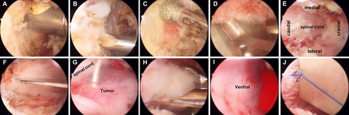
Figure 3 (A and B) The incisions and the resected tumor. (C) Hematoxylin and eosin (H&E) staining of the surgical biopsy specimen indicated that the tumor cells were arranged in bundles and swirls with gravel bodies evident, and their nuclei were ovoid or spindle-shaped.
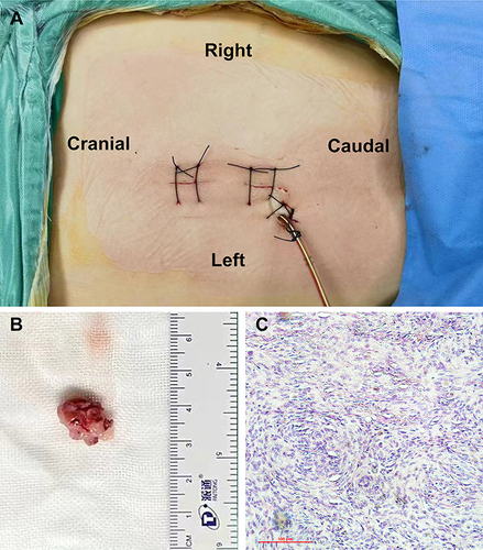
Figure 4 Computed tomography scan (CT) of post-operation. (A) Postoperative coronal CT of thoracic spine. (B) Postoperative sagittal postoperative CT. (C) Postoperative 3-dimensional CT. (D and E) Postoperative axial CT. Red arrow: the bone window.
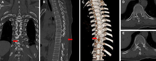
Figure 5 MRI sagittal (A–C) and axial (D and E) of post-operation showed that the IDEM tumor was removed completely, and the spinal cord returned to a normal position. The red and yellow lines represent the axial planes in (C).