
Figures & data
Point your SmartPhone at the code above. If you have a QR code reader the video abstract will appear. Or use:
Figure 1 The T2-weighted magnetic resonance imaging (MRI) radiographs are shown in sagittal (A) and axial view (B). The tumor compresses the nerve root to cause right L5 radiculopathy (arrow).
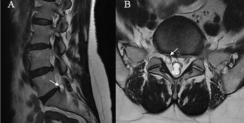
Figure 2 The right L5 root tumor is exposed. A number 11 sharp knife was used to make a small hole to open the dura.
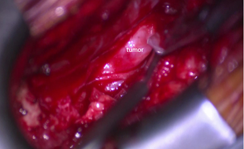
Figure 3 The lower margin of the tumor compared to normal nerve tissue.
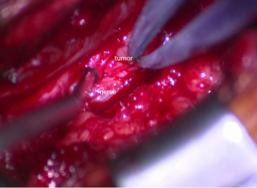
Figure 4 A micro-forceps was used to remove the tumor piecemeal.
