Figures & data
Figure 1 Pathologic macroscopic image of the tumor surgically excised. Measuring 1.7×1.2 x 1.2 cm.
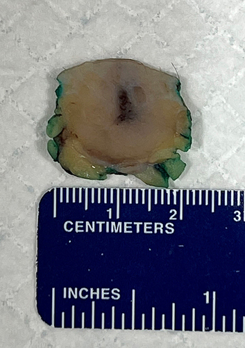
Figure 2 Higher magnification of the tumor cells highlights the cytologic features of enlarged nuclei, nuclear molding, and scant cytoplasm. Numerous mitotic figures and apoptotic cells are seen. (Hematoxylin-eosin, original magnification x20).
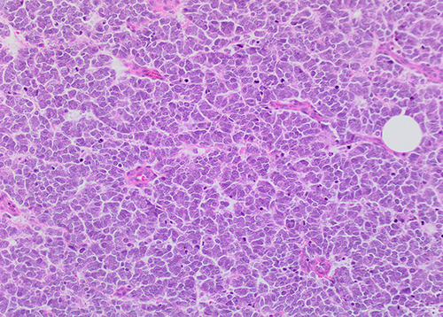
Figure 3 Tumor cells show positive, dot-like, staining for cytokeratin 20. (Immunohistochemical stain, original magnification x20).
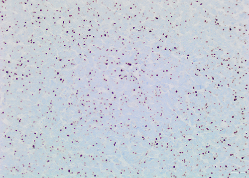
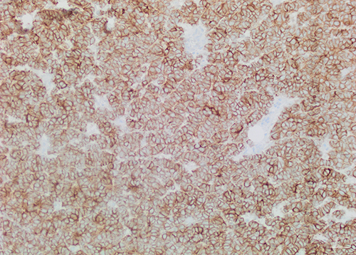
Figure 4 Tumor cells show diffuse positive staining for CD56. (Immunohistochemical stain, original magnification x20).