Figures & data
Figure 1 Light microscopy findings (×200). (a) Hematoxylin-eosin and (b) Periodic acid-Schiff-stained specimens show normal glomeruli and tubulointerstitial damage with tubular lymphocytic infiltration. (c and d) Electron microscopy findings. Normal glomeruli and nuclear and vacuolar degeneration in the tubules are observed (white arrows).
Notes: ▲: cytoplasmic vacuolar degeneration of the tubular epithelium. Δ: necrotic debris of the tubular lumen. ➜: tubulointerstitial inflammation.
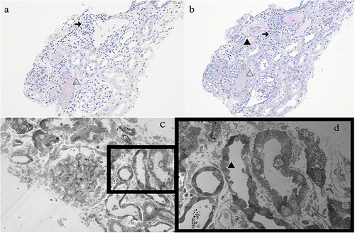
Figure 2 Light microscopy findings (×200). Hematoxylin-eosin–stained specimen shows chorionic villi without enlargement and decidua with hemorrhagic changes.
Note: ➜: chorionic villi without enlargement.
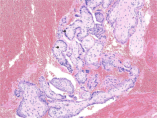
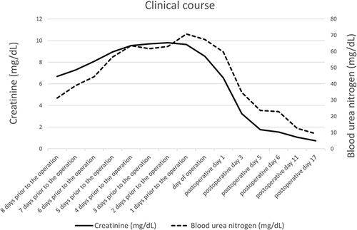
Figure 3 Blood urea nitrogen and serum creatinine levels during the peri-operative period.