Figures & data
Figure 1 Orbital photograph showing proptosis, exophthalmic and edema with hyperemia.
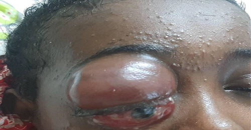
Figure 2 Orbital MRI showed space-occupying that’s T1-isointense and T2-hyperintense heterogeneous lobulated lesion with central signal void in the right intraorbital region resulting exophthalmos.
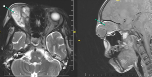
Figure 3 DWI and ADC MRI: Lesions showing acute diffusion restriction were detected in both parietal, frontal and right occipital lobes (acute/subacute ischemic infarct).
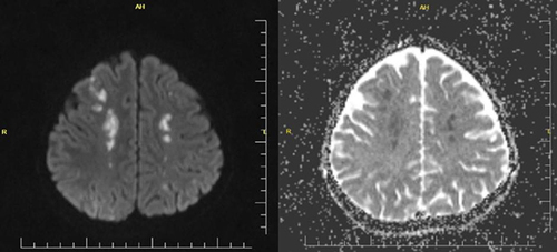
Figure 4 The smear shows fungal hyphae with broad based non-septate hyphae stained with H&E stain.
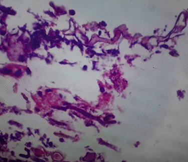
Figure 5 Day 6 photo taken following the initiation of antifungal therapy.
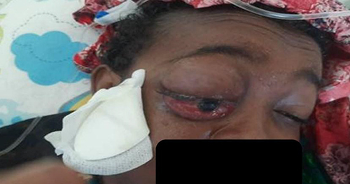
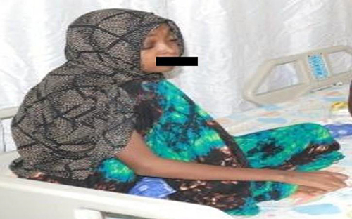
Figure 6 The final day of the hospital discharge.