Figures & data
Figure 1 Brain MRI images: Axial T2WI (A), FLAIR (B), DWI (C and D), and Apparent Diffusion Coefficient (E and F) map of a patient’s brain reveal acute infarction in the left middle cerebral artery territory. The scans show significant hyperintensity in the left insular cortex, body of the caudate nucleus, posterior part of the lentiform nucleus, and lateral part of the inferior frontal lobe, with evident restricted diffusion (white arrow). The gradient echo image (G) displays a blooming artifact in the M2 segment of the left middle cerebral artery (black arrow), likely indicating a thrombosed vessel.
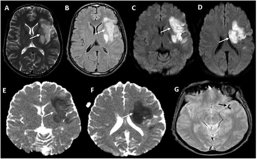
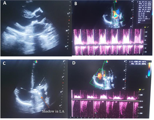
Figure 2 Transthoracic echocardiography showing a well seated mitral metallic valve (A) and normal trans mitral prosthetic valve mean pressure gradient of 3.7 mmHg (B), Apical 4-chamber view showing a metallic prosthetic valve at the mitral valve area (C) (white arrow) and mechanical valve shadow in the left atrium (LA) (orange arrow) with normal velocity and pressure gradient across the tricuspid valve (D).