Figures & data
Figure 1 (a) Coronal contrast-enhanced CT scan of the brain reveals a filling defect along the superior sagittal sinus, presenting as a delta sign (indicated by an arrow). (b) Sagittal contrast-enhanced CT scan of the brain displays a tubular filling defect along the posterior aspect of the superior sagittal sinus (highlighted by an arrow).
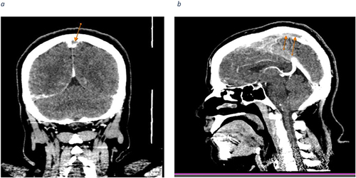
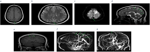
Figure 2 (a) Axial T1WI-SE brain MRI shows slightly high signal intensity of superior sagittal sinus posteriorly (arrow) compared to cerebral white matter. (b) and (c) Axial T2-weighted and Diffusion-weighted Imaging (DWI) Brain MRI depicting high signal intensity along the posterior aspect of the superior sagittal sinus (indicated by arrows) respectively. (d) and (e) Sagittal T2-weighted spin echo (T2WI- SE) and coronal T1-weighted spin echo (T1WI-SE) brain MRI demonstrating hyperintensity along the posterior aspect of the superior sagittal sinus and the right vein of Trolard (superior anastomotic vein) respectively. (f) and (g) The MRV reveals a focal absence of blood flow (filling defect) along the posterior portion of the superior sagittal sinus and the right vein of Trolard (superior anastomotic vein).