Figures & data
Figure 1 Under 10X magnification, shows a hemorrhagic background containing cohesive clusters of slender spindle cells with a taper-ended nucleus set in a pinkish stromal fragment, along with small mature lymphocytes and plasma cells in the background.
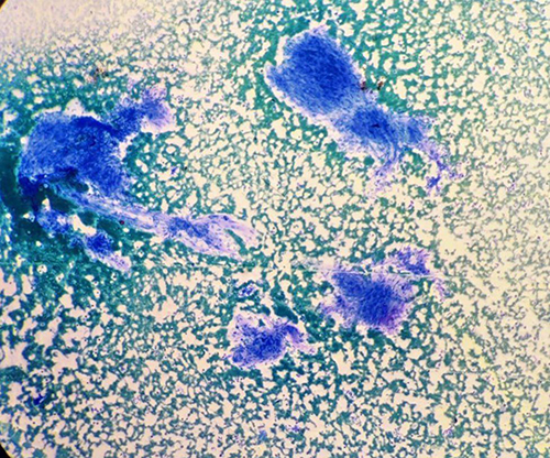
Figure 2 Under10X magnification, shows a hemorrhagic background containing single cells and cohesive clusters of slender spindle cells with a tapper-ended nucleus, along with small mature lymphocytes and plasma cells in the background.
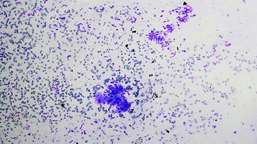
Figure 3 Under 100X magnifications, shows a hemorrhagic background containing a cohesive cluster of slender spindle cells exhibiting mild to moderate pleomorphism along with small mature lymphocytes and plasma cells at the background set in pinkish stroma.
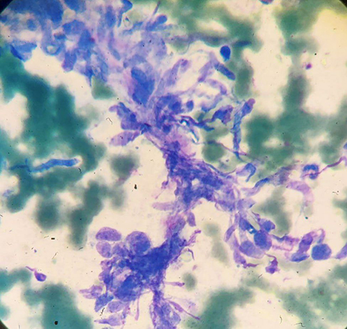
Figure 4 Under10X magnification, shows a hemorrhagic background containing a cohesive cluster of slender spindle cells set in a pinkish stroma.
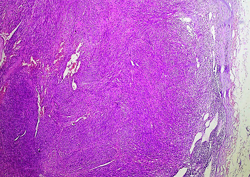
Figure 5 Under 20X magnifications, shows lymphoid tissue with a completely effaced architecture composed of vague interlacing fascicles of spindle cells along with extravasated RBCs. Subcapsular remnants of lymphoid aggregates are also noted.
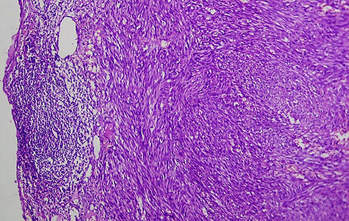
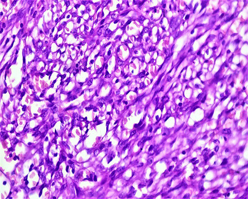
Figure 6 Under 100X magnifications, shows vague interlacing fascicles of hyperchromatic and pleomorphic spindle cells forming vascular slit-like and blood-filled spaces. Accompanying mononuclear inflammatory infiltrates and occasional mitotic figures, including abnormal forms, are seen.
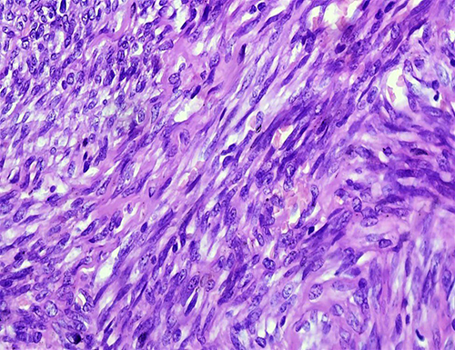
Figure 7 Under 100X magnifications, shows hyperchromatic spindle cells forming vascular slits and sieve-like blood-filled spaces. Accompanying mononuclear inflammatory infiltrates.