Figures & data
Figure 1 This is a patient’s image showing the left fronto-maxillary reddish-purple patch, which indicated port-wine of the face.
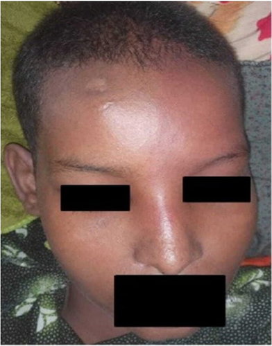
Figure 2 A computed tomography of the brain reveals unilateral left-sided moderate atrophy of the cerebral hemisphere (A) with extensive gyriform cortical-subcortical (tram-track) calcifications (B).
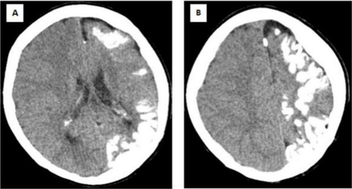
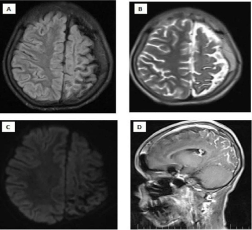
Figure 3 Magnetic Resonance imaging of the brain which shows left cerebral hemisphere, prominent atrophic changes at the sulcus and fissures of the parietal lobe and thickening of the adjacent of the left parietal lobe are noteworthy (A and B). There is no diffusion restriction (C). After Intravenous contrast injection (D). There is gyral-meningeal contrast enhancement in the cortical regions in the areas described of the left parietal lobe.