
Figures & data
Figure 1 Patient 1’s AS-OCT, OCT, and Fundus results at 92 days after microhook surgery. (A) The AS-OCT revealed ciliary detachment on both the nasal and temporal sides. The temporal ciliary detachment was larger. The anterior chamber was very narrow. (B) The OCT sliced vertically on the macular area. Retinal folds were observed. (C) Fundus results: retinal folds due to low IOP were observed. (D) Ultrasound biomicroscopy (UMB) revealed ciliary detachment on both the nasal and temporal sides.
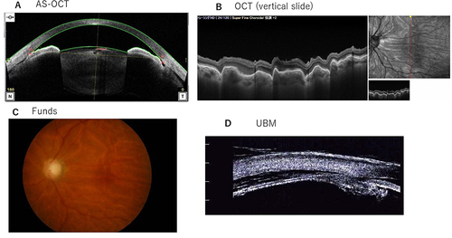
Figure 2 Patient 1’s AS-OCT, OCT, and Fundus results at 36 days after SF6 gas tamponade treatment. (A) The AS-OCT demonstrated that the ciliary body was attached. (B) As shown by OCT, retinal folds were improved. (C) The Fundus results revealed that the retinal folds were improved.
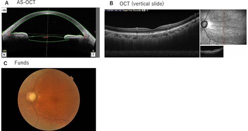
Figure 3 Patient 2’s AS-OCT, OCT, and Fundus results at 41 days after microhook surgery. (A) As shown by AS-OCT, ciliary detachment was present on both the nasal and temporal sides. The nasal ciliary detachment was larger. (B) OCT sliced vertically on the macular area. Radial folds around the fovea as hypotonic maculopathy were observed. (C) Fundus results: chorioretinal folds characteristic of hypotonic maculopathy were observed.
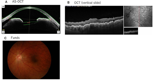
Table 1 Summary of Patient 1
Table 2 Summary of Patient 2
Figure 4 Patient 2’s AS-OCT results at 19 days after SF6 gas tamponade treatment. The ciliary detachment had improved, but the ciliary body did not attach.
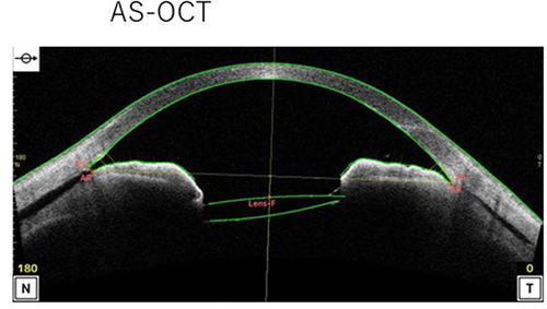
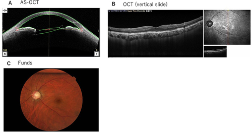
Figure 5 Patient 2’s AS-OCT results at 11 days after C3F8 gas tamponade treatment. (A) The AS-OCT demonstrated that the ciliary body was attached. (B and C) OCT and Fundus results at 55 days after the C3F8 gas tamponade treatment. (B) OCT showed that the retinal folds were improved. (C) Fundus results: approx. 20% of the C3F8 gas remained in the vitreous cavity. The retinal folds were improved.
Data Sharing Statement
The datasets in our study are available from the corresponding author on reasonable request.