Figures & data
Table 1 Hematology Examination Result
Figure 1 Haemorrhagic crusts on the lips, along with multiple ulcerations on the oral mucosa (A–C, E-H and J); Palatal erythema (D); Pseudomembranous candidiasis can be seen on the patient’s tongue (I).
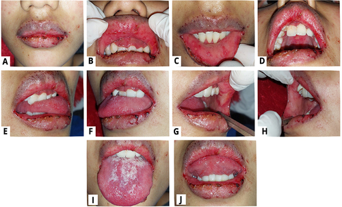
Figure 2 Overall improvements in the patient’s oral condition on the second visit (A–I).
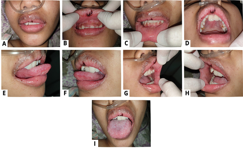
Figure 3 Improvement of the oral lesion on the 10th day (A–K).
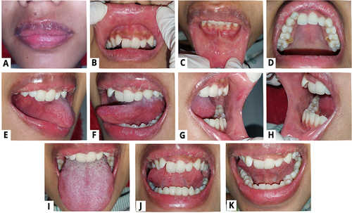
Figure 4 Patient has no complaints in the oral cavity (A–K).
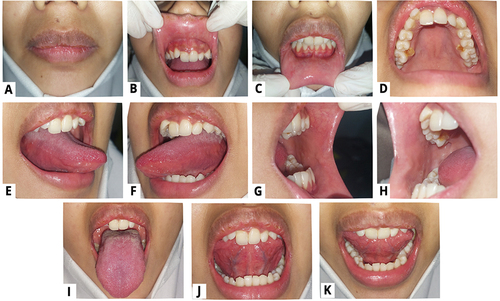
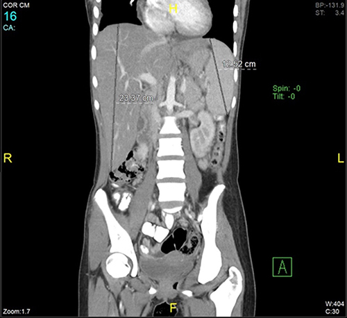
Figure 5 The hepatosplenomegaly condition can be seen on CT scan image.