Figures & data
Figure 1 Abdominal plain X-rays showing an oval calcific opacity.
Notes: Abdominal plain X-rays showing an oval calcific opacity (arrow) (A) in the left pelvic area, (B) which migrated toward the midline in the next abdominal plain X-ray obtained after a week.
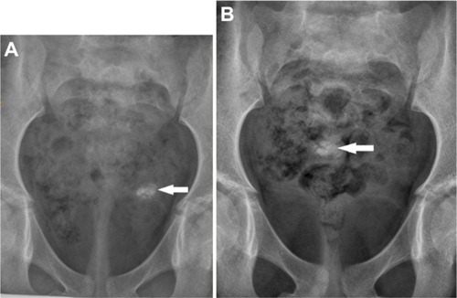
Figure 2 Axial computed tomography scan (bone window) image showing oval calcification (arrow) in the right adnexa.
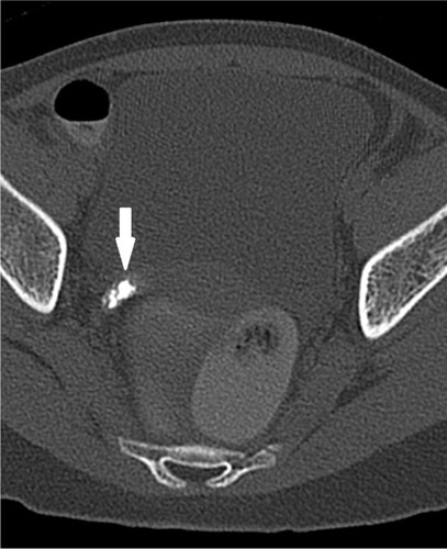
Figure 3 Axial short TI inversion recovery magnetic resonance image showing absent right adnexa and blunt ending right fallopian tube.
Note: Blunt ending right fallopian tube (black arrow); normal left ovary is noted (white arrow).
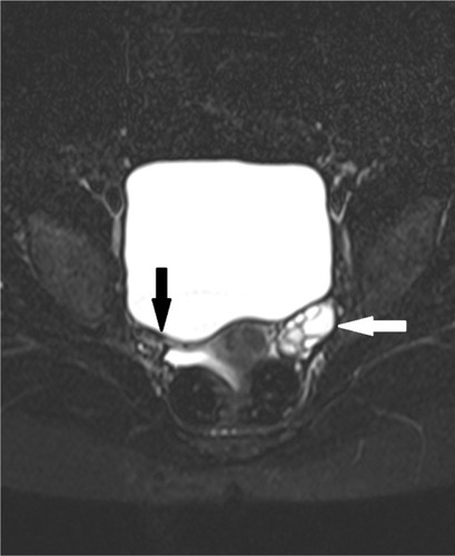
Figure 4 Axial fat suppressed T1-weighted image showing oval hyperintense lesion with thick central calcification in the left adnexa (arrow).
Note: Peripheral hyperintensity on the T1-weighted image can be due to methemoglobin or calcification.
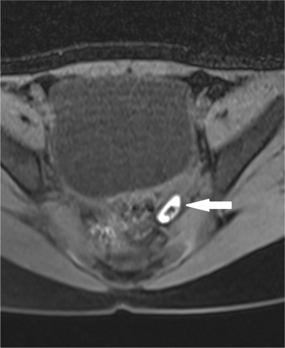
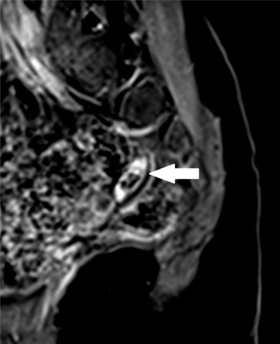
Figure 5 Sagittal post-gadolinium contrast T1-weighted fat suppressed image shows nonenhancing calcified oval lesion (arrow) in the left adnexa.