Figures & data
Figure 1 Frontal chest radiograph.
Notes: This frontal chest radiograph demonstrates surgical emphysema along the upper part of the right lateral chest wall and the right side of the neck (black arrow), air within the superior mediastinum on the right (white arrow), and the loss of volume of the right lung leading to a mediastinal shift to the right and the elevation of the right diaphragmatic dome.
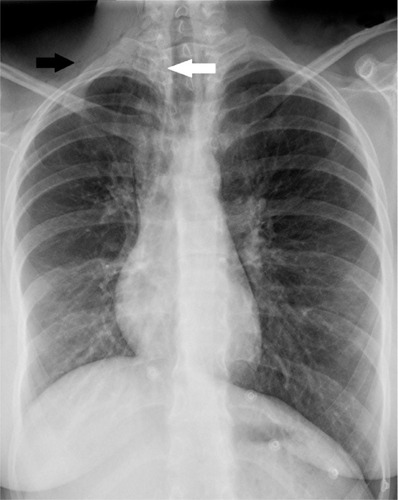
Figure 2 Portable bedside semi-sitting chest radiograph.
Notes: This radiograph shows the increased severity of pneumomediastinum (white arrow), surgical emphysema (black arrow), right lung collapse, and mediastinal shift to the right.
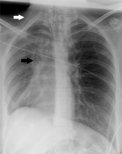
Figure 3 Axial nonenhanced chest CT scan.
Notes: This chest scan shows air dissecting through the mediastinal spaces (down arrow) and the subcutaneous soft tissue (up arrow). Minimal pneumothorax is also noted (left directional arrow).
Abbreviation: CT, computed tomography.
Abbreviation: CT, computed tomography.
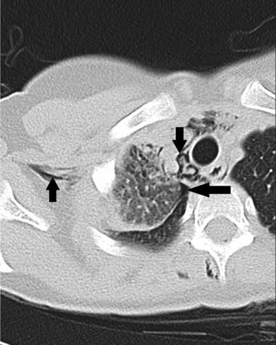
Figure 4 Axial nonenhanced chest CT scan.
Notes: This chest CT scan shows air dissecting through the mediastinal spaces (down arrow) and the subcutaneous soft tissue (right directional arrow). Air is also noted within the extradural space in the lower cervical and the upper thoracic spine (up arrow).
Abbreviation: CT, computed tomography.
Abbreviation: CT, computed tomography.
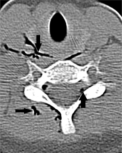
Figure 5 Sagittal reformatted CT scan images of chest.
Notes: This chest scan demonstrates air within the extradural space in the lower cervical and upper thoracic spine (arrows).
Abbreviation: CT, computed tomography.
Abbreviation: CT, computed tomography.
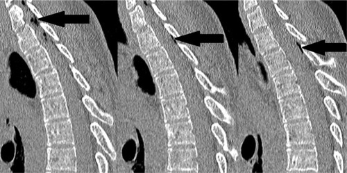
Figure 6 Coronal and axial reformatted CT scan images of the chest.
Notes: (A) This CT scan image demonstrates surgical emphysema along the upper part of the right lateral chest wall and the right side of neck (down arrow), air within the superior mediastinum on right (right directional arrow), loss of volume of the right lung leading to the mediastinal shift to the right, elevation of the right diaphragmatic dome, and obstruction of the segmental right upper lobe bronchus by mucus plugs (A and B) (up arrows).
Abbreviation: CT, computed tomography.
Abbreviation: CT, computed tomography.
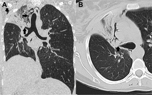
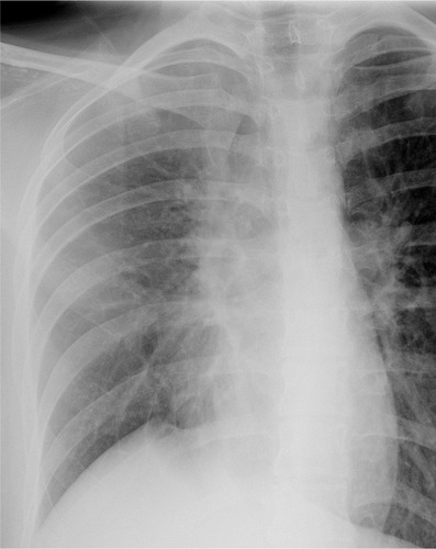
Figure 7 Follow-up frontal chest radiograph.
Notes: This chest radiograph demonstrates the complete resolution of surgical emphysema and pneumomediastinum and significant inflation of the right lung on the fifth hospital admission day.