Figures & data
Figure 1 Unenhanced axial T2-weighted (A and B), fluid attenuated inversion recovery (C and D), and T2*-weighted (E and F) images showing hyperintensities involving the cerebral and cerebellar cortex and white matter, basal ganglia, thalami, and brainstem bilaterally (solid black arrows). A focal hyperintense lesion is noted in the right temporal periventricular white matter (open black arrow). Petechial hemorrhages are noted in the brainstem and gangliocapsular regions bilaterally (white arrows).
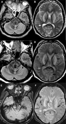
Figure 2 Unenhanced T2-weighted coronal (A) and sagittal (B) images of the brain and cervical spinal cord (C) showing hyperintensities involving the cerebellar cortex and brainstem (solid black arrows). A focal hyperintense lesion is noted in the right temporal periventricular white matter (solid white arrow). Petechial hemorrhages are noted in the brainstem (open black arrows). The cervical spinal cord is swollen and reveals hyperintense signals within it (open white arrow).
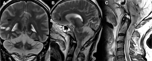
Figure 3 Axial diffusion-weighted image showing restricted diffusion involving the entire cerebellum bilaterally (arrows).
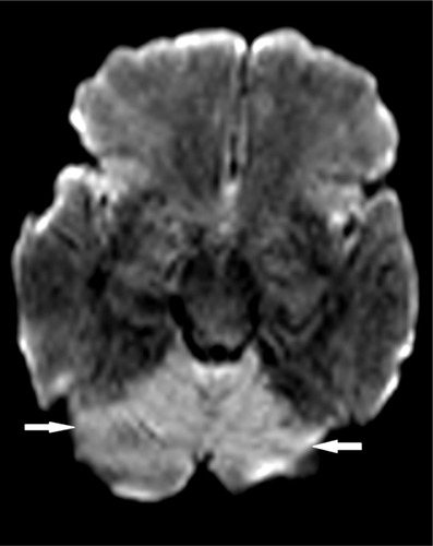
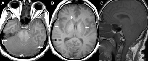
Figure 4 Unenhanced T1-weighted axial images of brain (A and B) and sagittal image of brain and cervical spinal cord (C) showing hypointensities involving cerebral and cerebellar cortex and white matter, basal ganglia, thalami, and brainstem bilaterally (solid white arrows). A focal hypointense lesion is noted in the right temporal periventricular white matter (black arrow). The cervical spinal cord is swollen and reveals hypointense signals within it (open white arrows).