
Figures & data
Figure 1 Preoperative pure tone audiogram showing a left-sided mixed hearing loss. Postoperative pure tone audiogram showing marked improvement, especially in the lower frequencies.
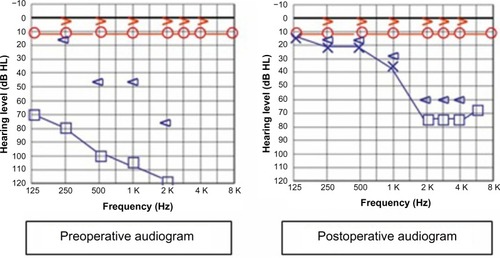
Figure 2 Axial computed tomography scan of the temporal bone showing air within the left vestibule (arrow).
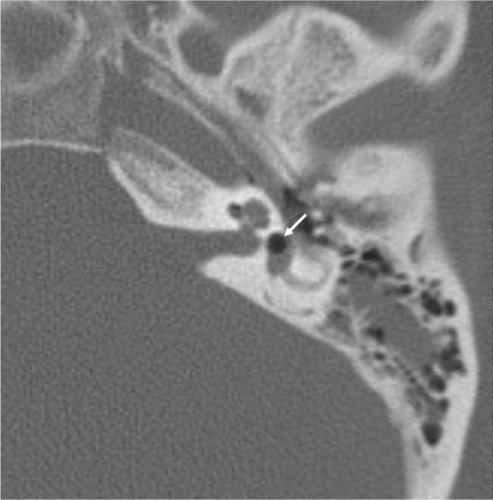
Figure 3 Coronal computed tomography scan of the temporal bone showing air within the basal turn of the cochlea (arrow).
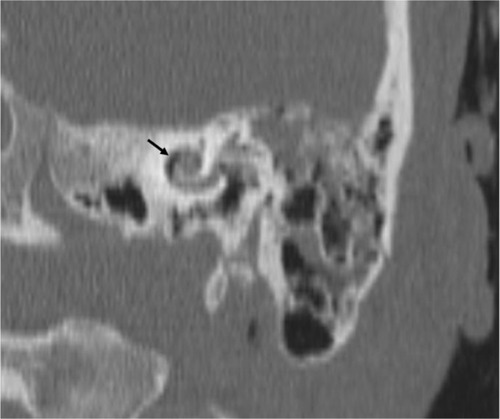
Table 1 Review of the literature