Figures & data
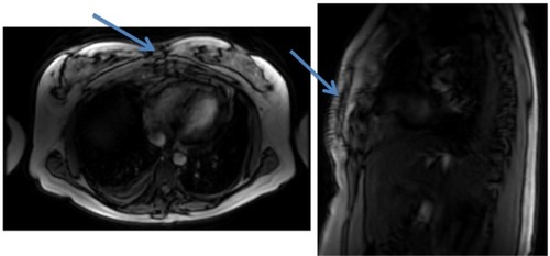
Figure 1 Electrocardiogram.
Note: This was a normal electrocardiogram with no changes.
Abbreviations: a, augmented; V, vector; L, left; R, right; F, foot.
Abbreviations: a, augmented; V, vector; L, left; R, right; F, foot.
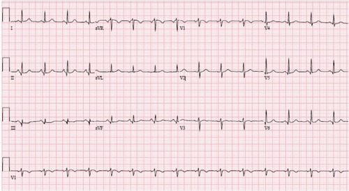
Figure 2 Chest X-rays.
Note: Chest X-rays show an interval development of a right pleural effusion that is best seen on the lateral view (arrow).
Abbreviation: L, left.
Abbreviation: L, left.
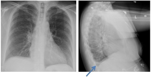
Figure 3 Magnetic resonance imaging of the chest.
Notes: Increased signal is seen on T2-weighted images in subcutaneous fat in the anterior abdominal wall at the midline. Increased signal is also noted in anterior mediastinum fat posterior to the sternum. There is periosteum enhancement of the sternum manubrium and at the upper part of the body of the sternum after contrast administration. There is also increased signal in the bone marrow noted on T2-weighted images in the sternum manubrium and in the upper part of the body of the sternum, suggesting bone marrow edema. No evidence of fracture is noted. Findings are consistent with osteomyelitis involving the manubrium and the upper part of the sternum body. Arrows indicate the area of osteomyelitis.