Figures & data
Figure 1 Pretreatment MRI.
Notes: (A–D, Top) Findings in brain images (images A, B FLAIR axial sequences and images C, D FLAIR sagittal sequences) are typical for primary demyelinating disease with several callosal plaques. (A–B, Bottom) Thoracic images (image A, T2 sagittal and image B, T2 axial), suggest possible demyelinating plaque at T3–4.
Abbreviations: MRI, magnetic resonance imaging; FLAIR, fluid-attenuated inversion recovery.
Abbreviations: MRI, magnetic resonance imaging; FLAIR, fluid-attenuated inversion recovery.
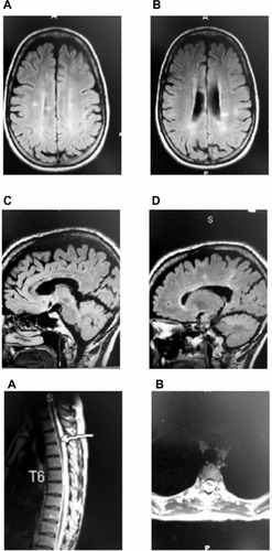
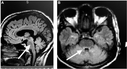
Figure 2 MRI following treatment with IVMP for 5 days.
Notes: T2 sagittal (image A) and axial (images B, C) images and T1 with gadolinium (image D) show new dorsal pontine white matter lesion as depicted by the arrows in image A and image B, located slightly eccentric to the right. Images do not show abnormal enhancement. No other changes were seen compared with pretreatment.
Abbreviations: IVMP, intravenous methylprednisolone; MRI, magnetic resonance imaging.
Abbreviations: IVMP, intravenous methylprednisolone; MRI, magnetic resonance imaging.
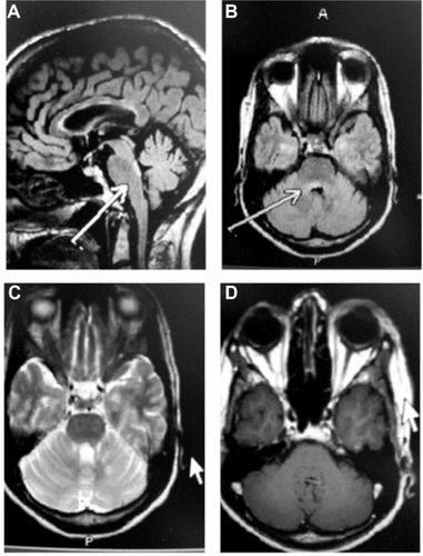
Figure 3 MRI 5 months following treatment with ACTH gel for 5 days, followed by monthly IV natalizumab.
Notes: Images are compatible with the patient’s known history of demyelinating disease. The lesion in the dorsal pons, shown by the arrows in image A (FLAIR sagittal) and image B (FLAIR axial), is less prominent than was seen prior to treatment with ACTH gel and IV natalizumab.
Abbreviations: ACTH, adrenocorticotropic hormone; IV, intravenous; MRI, magnetic resonance imaging; FLAIR, fluid-attenuated inversion recovery.
Abbreviations: ACTH, adrenocorticotropic hormone; IV, intravenous; MRI, magnetic resonance imaging; FLAIR, fluid-attenuated inversion recovery.