Figures & data
Figure 1 Frontal view of fractured mandibular teeth.

Figure 2 Occlusal view of fractured teeth. Glass Ionomer Cement is covering the fractured teeth.
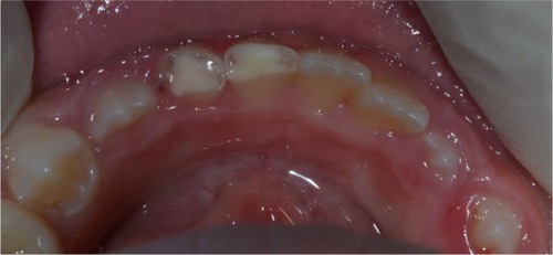
Figure 3 Fractured fragments after cleaning.
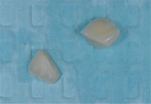
Figure 4 Circumferential enamel bevel placed on both fractured teeth.
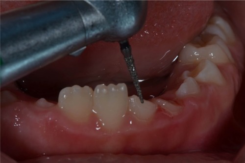
Figure 5 Fractured fragments received a circumferential enamel bevel.
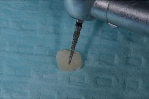
Figure 6 Frontal view showing excellent esthetics of attached fragments.
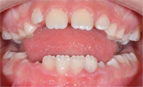
Figure 7 Frontal view of teeth after 8 month recall.
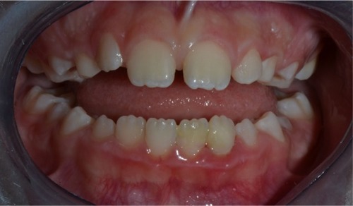
Figure 8 Lingual view showing the retainer in place. Some plaque accumulation noticed.
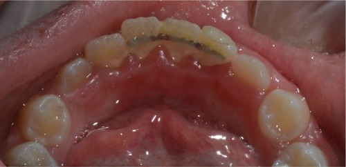
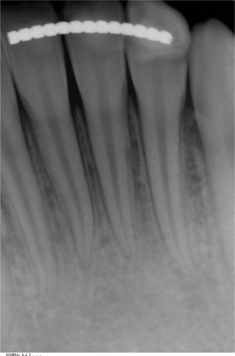
Figure 9 Periapical radiograph of mandibular incisors showing healthy pulp and periodontal tissues.