Figures & data
Figure 1 External examination of extraocular eye movements.
Notes: Images demonstrating complete ophthalmoplegia of the right eye with ptosis prior to surgery or amphotericin B treatment (left), followed by images obtained after 10 months showing full levator function with restoration of right extraocular eye movement in each position of gaze (right).
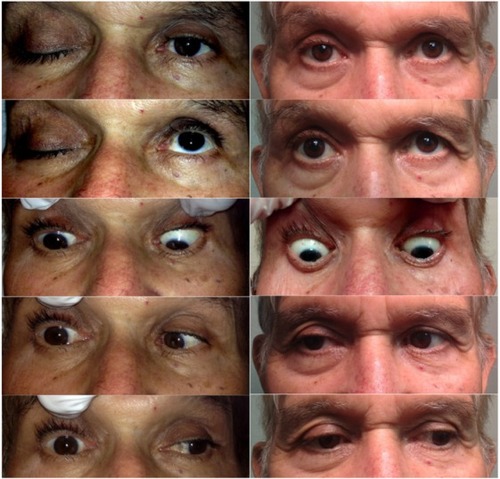
Figure 2 CT scan of the brain, without contrast.
Notes: CT scan displaying bilateral opacification of the ethmoids, near complete opacification of the right sphenoid, and mild mucosal thickening in the left sphenoid and maxillary sinuses with extension into the orbit. The coronal sectional image illustrates the proximity of the lesion next to the foramen rotundum and optic nerve foramen (A). The axial cross section discloses the opacification being significantly more affected on the right side of the sphenoid and ethmoid sinus, with some thickening on the left sphenoid sinus region. There is also involvement of the right middle and posterior orbital cavity adjacent to the optic foramen (B). The sagittal section illustrates the opacity reaching back toward the naropharynx. The inferior, middle, and superior turbinates are also affected, with some suggestion of involvement of the cryptoform plate (C).
Abbreviation: CT, computed tomography.
Abbreviation: CT, computed tomography.
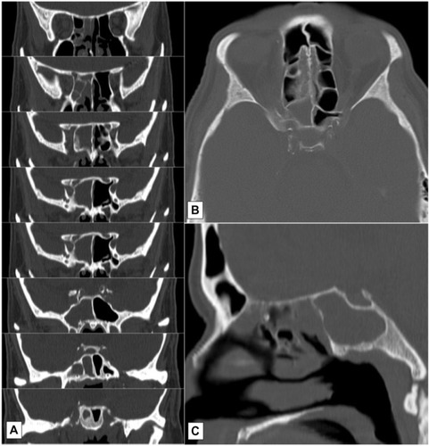
Figure 3 Histopathology slides.
Notes: Hematoxylin and eosin stain (200×) obtained from mucosal tissue of the ethmoid sinus showing necrotic and edematous tissue with neutrophilic infiltrate. There are also hyphae noted in this section, indicated by the arrows (A). Two Gömöri methenamine silver stains (400×) from the same biopsy showing broad, aseptate hyphae with right angle branching, also indicated by the arrows (B and C).
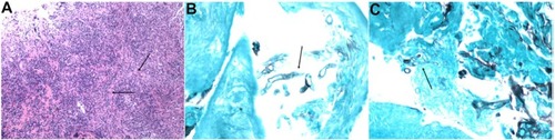
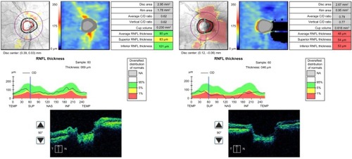
Figure 4 Optical coherence tomography.
Notes: Cirrus-HD optical coherence tomography (Carl Zeiss Meditec, Inc., Oberkochen, Germany) of the right optic nerve at initial presentation (left), followed by an additional scan obtained 10 months later demonstrating enlargement of the physiologic cup and generalized loss of retinal nerve fiber displaying gradual presynaptic atrophy of the right optic nerve (right).
Abbreviations: RNFL, retinal nerve fiber layer; C/D, cup to disc; T/TEMP, temporal; SUP, superior; N/NAS, nasal; INF, inferior; HD, high definition; OD, right eye; NA, not available.
Abbreviations: RNFL, retinal nerve fiber layer; C/D, cup to disc; T/TEMP, temporal; SUP, superior; N/NAS, nasal; INF, inferior; HD, high definition; OD, right eye; NA, not available.