Figures & data
Figure 1 Slit-lamp photograph of the right eye.
Notes: (A) Slit-lamp photograph of the right eye. (B) Descemet’s scroll, indicated by the arrowheads. (C) The Descemet’s scroll appears as a translucent rod with a central core and (D) it extends into the anterior chamber.
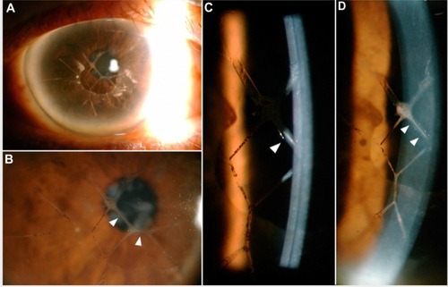
Figure 2 Slit-lamp photograph of the left eye.
Note: (A) Slit-lamp photograph of the left eye (B) showing a fine Descemet’s scroll and (C) stromal ghost vessels.
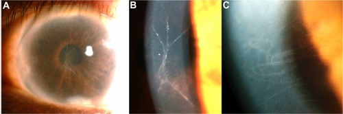
Figure 3 Slit-lamp photograph.
Note: (A) Arrowhead shows pupillary membrane in the right eye and (B) Arrowheads show Koeppe nodules in the left eye.
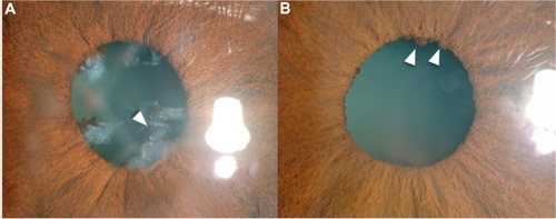
Figure 4 OD and OS confocal microscopy show endothelial pleomorphism and polymegathism (40×).
Abbreviations: OD, right eye; OS, left eye.
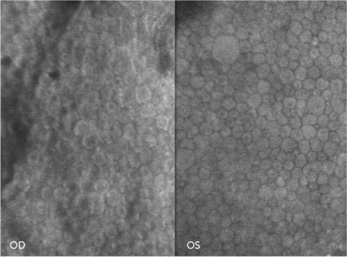
Figure 5 Confocal microscopy of the right eye shows a Descemet’s scroll.
Notes: The scrolls appeared as (A and B) tubular structures with (C) outer hyperreflectivity and inner hyporeflectivity. (D) Some areas show cellular components on the surface.
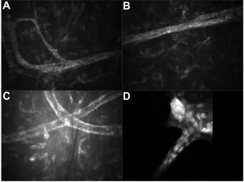
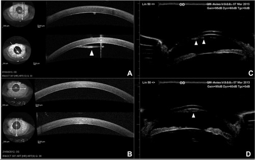
Figure 6 OD and OS AS-OCT and OD ultrasound biomicroscope.
Notes: (A) OD and (B) OS AS-OCT show a thickened Descemet’s membrane; (A) the scroll extended from the posterior corneal surface into the anterior chamber. Arrowhead shows the rod-shaped Descemet’s scroll. (C and D) UBM of the right eye shows Descemet’s scrolls. Arrowheads show the rod-shaped Descemet’s scroll.
Abbreviations: OD, right eye; OS, left eye; AS-OCT, anterior segment optical coherence tomography; UBM, ultrasound biomicroscopy.
Abbreviations: OD, right eye; OS, left eye; AS-OCT, anterior segment optical coherence tomography; UBM, ultrasound biomicroscopy.