Figures & data
Figure 1 The chest computed tomography showed a large anterior mediastinal cystic mass lateralized to the left side frame evoking a cystic teratoma or a cystic thymoma without aggressive loco-regional signs (arrow).
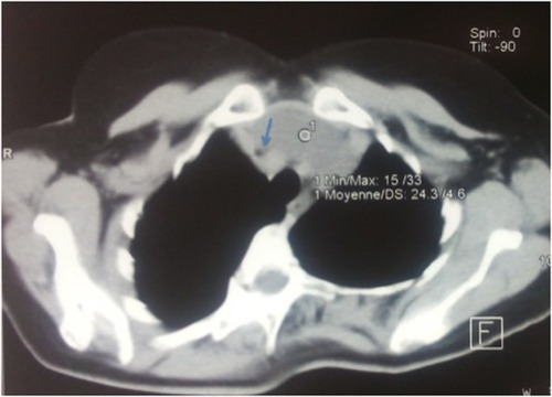
Figure 2 Gross examination showed a unilocular cystic mass (arrow) with some thymic lobules (star).
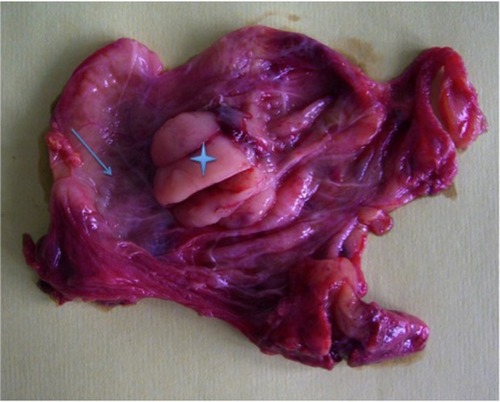
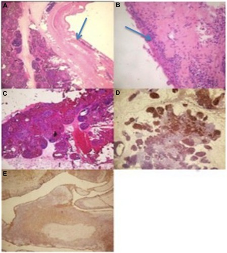
Figure 3 Microsopic findings.
Notes: (A) The coexistence of a cystic lesion (arrow) and true thymic hyperplasia (hematoxylin and eosin stain [HE] ×250; (B) the epithelial lining (arrow) of the cyst was mainly flattened and focally hyperplastic (HE ×400); (C) true thymic hyperplasia is characterized by the conservation of the thymic architecture which consists of a corticomedullary differentiation and the presence of many Hassall’s corpuscles in the medulla (HE ×250); (D) immunohistochemical study showing the positivity of the lymphocytes with the terminal deoxynucleotidyl transferase antibody (HE ×250); (E) immunohistochemical study showing the positivity of the epithelial lining with the cytokeratin antibody (HE ×250).