Figures & data
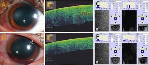
Figure 1 Marked focal corneal edema → marked focal stromal edema.
Notes: (A) Anterior segment photography showing a marked focal corneal edema. (B) Anterior segment optical coherence tomography demonstrating severe corneal swelling and markedly increased corneal thickness in the affected area. (C) Specular microscopy revealing decreased endothelial cell density and increased corneal thickness. (D) Anterior segment photography showing resolution of corneal edema through conservative management. (E) Anterior segment optical coherence tomography demonstrating complete resolution of corneal swelling. (F) Specular microscopy revealing substantially decreased endothelial cell density in the left (L) eye compared to the right (R) eye.