Figures & data
Figure 1 Clinical picture and H & E staining.
Notes: (A and B) Showing restriction of down gaze in right eye (A) and right orbital fullness (B). (C) Renal biopsy revealed increased endocapillary cellularity (yellow arrow), cellular crescent (black arrow), and chronic interstitial inflammation (red arrow); (H&E 10×).
Abbreviation: H&E, hematoxylin and eosin.
Abbreviation: H&E, hematoxylin and eosin.
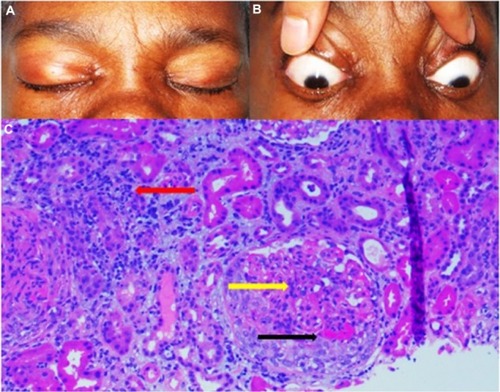
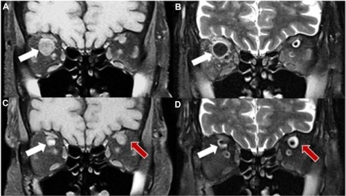
Figure 2 MRI of orbits.
Notes: (A and B) Showing coronal section (T1 and T2) at initial presentation. White arrow pointing SOVT in right eye. (C and D) Showing MRI of orbit 4 weeks later; coronal section (T1 and T2). White arrow pointing to resolving superior ophthalmic vein thrombosis in right eye. Red arrow pointing to new SOVT in left eye.
Abbreviation: SOVT, superior ophthalmic vein thrombosis; MRI, magnetic resonance imaging.
Abbreviation: SOVT, superior ophthalmic vein thrombosis; MRI, magnetic resonance imaging.