Figures & data
Figure 1 CT images of 52 year old male.
Notes: (A) Arterial phase image. The lesion in segment 7–8 has increased contrast enhancement according to liver parenchyma (white arrow). (B) Portal phase image. The lesion appears hypodense. The lesion in the left lobe has focal contrast material accumulation pooling and contrast output leakage outside the liver is visible (black arrowhead). (C) Venous phase image. Delayed phase reveals focal contrast material accumulation pooling in the large lesion of the left lobe (black arrow).
Abbreviation: CT, computed tomography.
Abbreviation: CT, computed tomography.
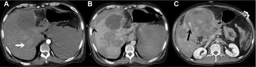
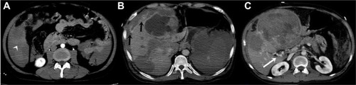
Figure 2 CT images of 52 year old male.
Notes: (A) Arterial phase image. The contrast accumulation in peritoneal cavity is prominent (white arrowhead) (B) Delayed phase image. The contrast accumulation outside of the liver is prominent (black arrows). (C) Portal phase image. Collateral vascular structures are visible at the hilus of the liver (white arrow).
Abbreviation: CT, computed tomography.
Abbreviation: CT, computed tomography.