Figures & data
Figure 1 Orbital depression due to orbital cavity emptiness in a newborn with severe right microphthalmia and left anophthalmia.
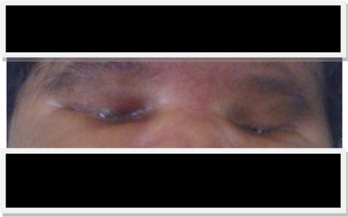
Figure 2 Ocular ultrasound of a newborn affected by (A) severe right-side microphthalmia with the presence of a vesicle in the right-side cavity, and (B) left-side anophthalmia with an empty orbital cavity.
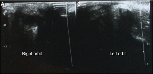
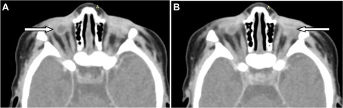
Figure 3 Axial section of ocular CT scan in parenchymal window after contrast injection.
Notes: (A) Right side microphthalmia; notice that the lens is absent, the optic nerve, the internal and external rectus muscles are present (arrow). (B) Left side anophthalmia; notice that there are no eye and no optic nerve. Internal and external rectus muscles are fixed in one place (arrow).
Abbreviation: computed tomography.
Abbreviation: computed tomography.