Figures & data
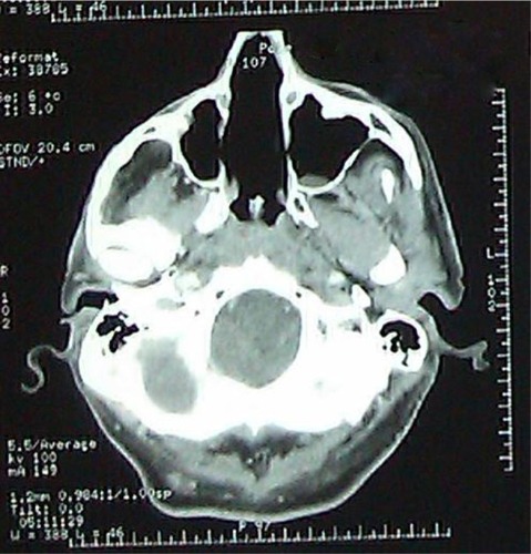
Figure 1 Preoperative CT scan showing the nasal tumor.
Notes: (A) Axial computed tomography scan of sinus with a parenchymal opening; (B) coronal bone with an opening showing a thickening tissue of the right nasal cavity.
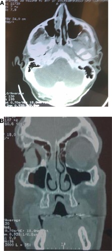
Figure 2 Infiltration of the right nasal cavity wall with a melanoma arriving in depth to the cartilage without invading it.
Notes: Hematoxylin-eosin-saffron staining (magnification × 2). Image courtesy of JL Kemeny.
Abbreviation: hematoxylin-eosin-saffron.
Abbreviation: hematoxylin-eosin-saffron.
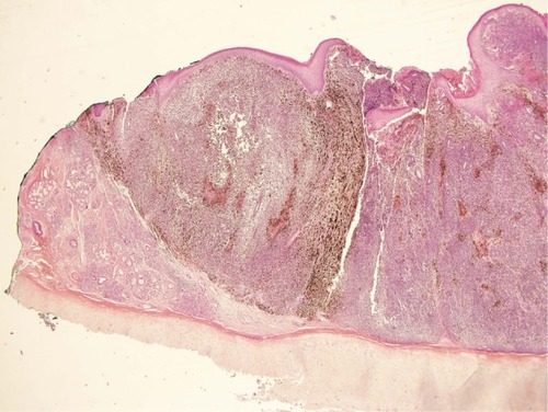
Figure 3 Detail of tumor melanoma cells that have an atypical nucleus. Note: Image courtesy of JL Kemeny.
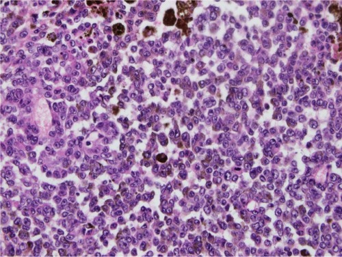
Figure 4 Brown marking of intracytoplasmic tumor cells with HMB45 and melan A in immunohistochemistry on paraffin section (magnification ×40).
Note: Image courtesy of JL Kemeny.
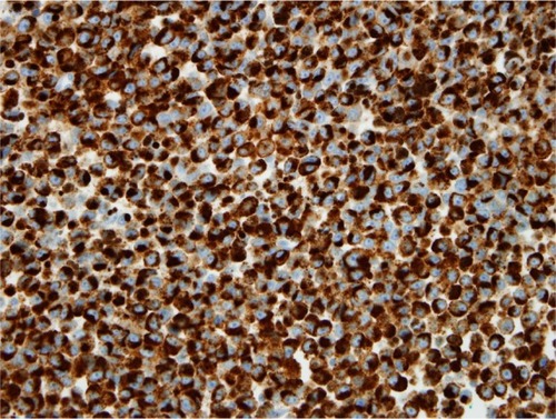
Figure 5 Postoperative computed tomography showing empty nasal cavities.