Figures & data
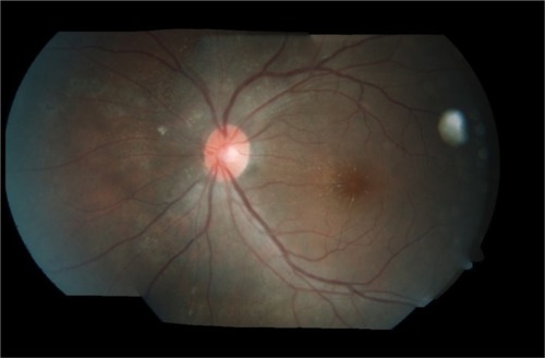
Figure 1 Left fundus showed swollen optic disc (blue arrow) with macular star (white arrow) and crops of active choroidal lesions at superonasal retina with a linear arrangement in the form of migratory track nasally (red arrow).
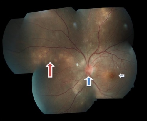
Figure 2 Optical coherence tomography of the left eye showed minimal subretinal fluid at the macula with some hyperreflectivity lesions at presentation. The scale represents the corresponding measurement of 200 mm vertically and horizontally.
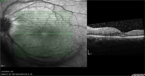
Figure 3 Left fundus showed resolution of crops of choroidal lesions with multiple focal laser scars nasal to the optic disc at 1 week posttreatment.
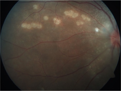
Figure 4 Optical coherence tomography of the left eye showed resolution of subretinal fluid at the macula at 1 week posttreatment. The scale represents the corresponding measurement of 200 mm vertically and horizontally.
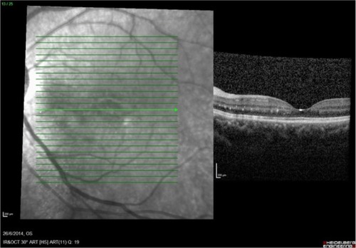
Figure 5 Left fundus showed regression of optic disc swelling with resolving macular star and resolution of crops of choroidal lesions with multiple focal laser scars nasal to the optic disc at 1 month posttreatment.