Figures & data
Figure 1 External appearance of a 15-year-old boy.
Note: NF1 skin findings, including café au lait spots and peripheral neurofibromas, are seen throughout his body.
Abbreviation: NF1, neurofibromatosis type 1.
Abbreviation: NF1, neurofibromatosis type 1.
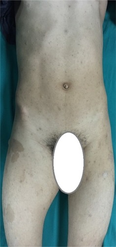
Figure 2 MRI appearance of a 13×9 cm mass at the anteromedial thigh.
Notes: (A) Isointense in T1W images, (B) heterogeneously iso- to hyperintense in T2W images, and (C) diffusely contrast enhancing except cystic areas, with posterior extension in postcontrast images.
Abbreviations: MRI, magnetic resonance imaging; T1W, T1 weighted; T2W, T2 weighted.
Abbreviations: MRI, magnetic resonance imaging; T1W, T1 weighted; T2W, T2 weighted.
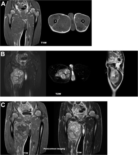
Figure 3 Macroscopic appearance of the mass after surgical excision.
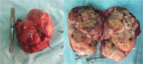
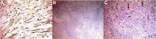
Figure 4 Appearance after histopathological and immunochemical staining.
Notes: (A) Immunohistochemical staining; S-100 positivity in tumor cells, (B) cellular tumoral area (H&E staining; original magnification: ×100), (C) pleomorphic (red arrows) and multinuclear (yellow arrows) tumor cells (H&E staining; original magnification: ×200).
Abbreviation: H&E, hematoxylin and eosin.
Abbreviation: H&E, hematoxylin and eosin.