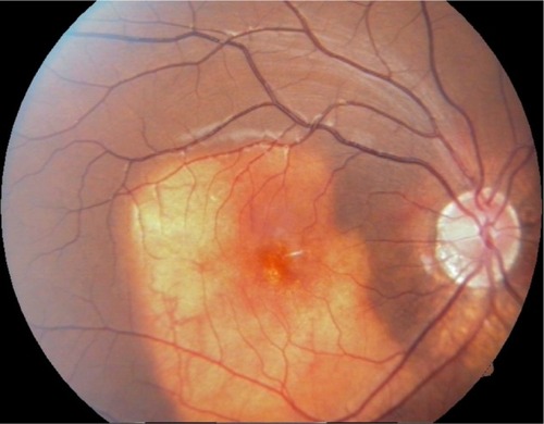
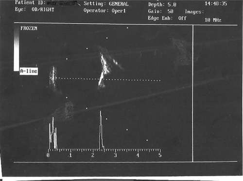
72
Views
3
CrossRef citations to date
0
Altmetric
Case Report

A case of choroidal osteoma in a 10-year-old child

&
Pages 273-275 | Published online: 02 Nov 2015

Figures & data

Figure 1 Fundus picture showing a typical orange-yellowish lesion.

Figure 1 Fundus picture showing a typical orange-yellowish lesion.

Figure 2 B-scan ultrasonogram showing hyper-reflectivity of the lesion persisting even at 60 dB gain.

Figure 2 B-scan ultrasonogram showing hyper-reflectivity of the lesion persisting even at 60 dB gain.