Figures & data
Figure 1 In 2008, Humphrey visual fields show left dense superior field loss with MD of −12.37 dB (A) and in 2010, progressive field loss with MD −14.25 dB threatening fixation is noted (B).
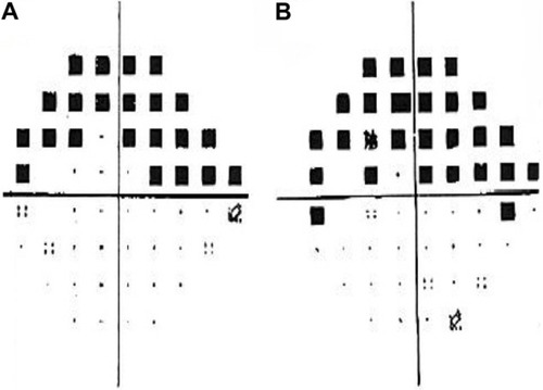
Figure 2 Four months following surgery, left eye anterior segment shows gold shunt in the superonasal quadrant in good position (A), with advanced cupping in a highly myopic left optic nerve (B).
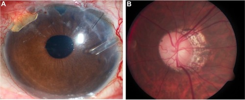
Figure 3 Four years following surgery, left eye anterior segment with gold shunt in good position (A), with stable glaucomatous optic neuropathy (B).

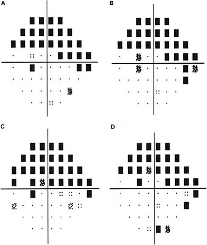
Figure 4 Humphrey visual fields show stable left visual fields following gold shunt surgery with MD of −11.84 dB at 1.5 years later (A), −12.21 dB at 2.5 years later (B), −14.20 dB at 3.5 years later (C), and −13.11 dB at 4.5 years later (D).