Figures & data
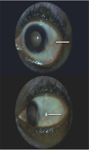
Figure 1 Right eye lesion before treatment with sulfasalazine.
Notes: White arrows show superior scleral node with mild perilesional inflammation and a quiet anterior chamber. Absence of epithelial defects (staining with fluorescein).
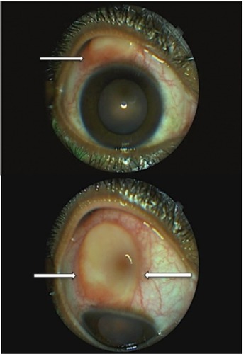
Figure 2 Left eye before treatment with sulfasalazine (use of fluorescein).
Note: White arrows show temporal corneal thickening and infiltrate (staining with fluorescein) associated with adjacent limbal infiltrate.
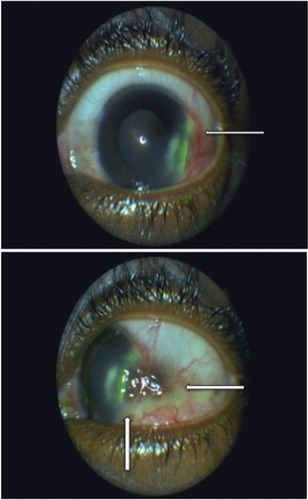
Figure 3 Left eye lesion 1 month after initiating the treatment with sulfasalazine.
Notes: White arrows show persistence of the temporal thinning of the cornea associated with a mild degeneration of the temporal conjunctiva. Disappearance of the infiltrate around the limbus.
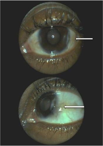
Figure 4 Right eye 8 months after initiating the treatment with sulfasalazine.
Note: The white arrow shows no reoccurrence of the scleritis in the superior quadrant.
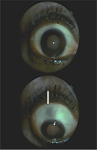
Figure 5 Left eye 8 months after initiating the treatment with sulfasalazine.
Note: White arrows show persistence of the temporal degeneration of the conjunctiva around the limbus that is growing on the cornea.