Figures & data
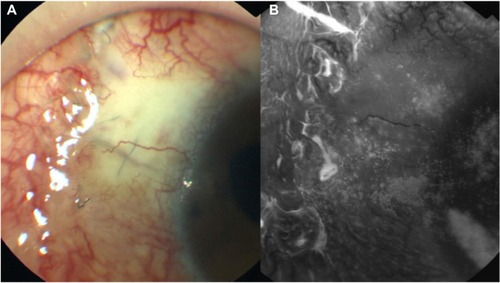
Figure 1 Apparent point leakage with severe ocular surface failure.
Notes: (A) Anterior segment photograph taken using a fundus camera 2 days after trabeculectomy. (B) The fundus camera in the fluorescein angiogram mode can distinguish exciting light from emitted light and allows the examination of epithelial failure in high contrast by using the appropriate filters. Severe epithelial failure of the bleb wall and cornea is observed and a leakage point is visible (arrows).
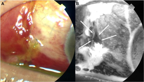
Figure 2 (A and B) Photographs obtained 2 days after the initiation of sodium hyaluronate eye drops.
Notes: The images show improvement of epithelial failure and resolution of the leakage point. Several microcysts are visible on the bleb surface at the sites where the epithelial failure showed improvement.
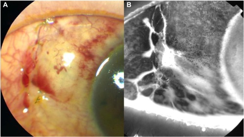
Figure 3 (A and B) Photograph taken 1 month after initiation of treatment with sodium hyaluronate eye drops.
Note: The microcysts are absent and the bleb surface is smooth.