
Figures & data
Figure 1 Tumor of entire left breast (rupture size 22×18 cm).
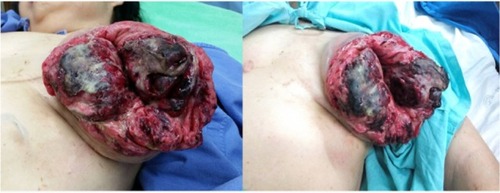
Figure 2 CT chest shows huge inhomogeneous enhanced mass at left breast.
Abbreviation: CT, computed tomography.
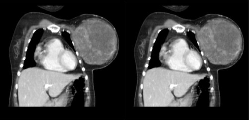
Figure 3 Microscopic study of phyllodes tumor.
Notes: Microscopic study of phyllodes tumor displays increased stromal cells with cleft- and leaf-like appearance of ducts and malignant spindle cells in the stroma with frequent mitoses (arrows) (H&E ×40).
Abbreviation: H&E, hematoxylin and eosin.
Abbreviation: H&E, hematoxylin and eosin.
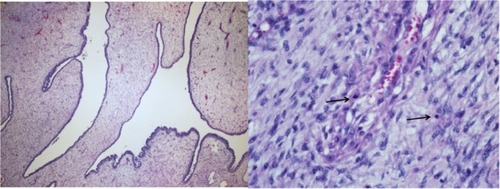
Table 1 Literature data of patients with ruptured phyllodes tumorCitation4,Citation6,Citation15,Citation16