Figures & data
Figure 1 Images illustrating the new AM-assisted TLE procedure.
Notes: (A) First, conventional TLE with a limbus-based conjunctival flap and a 3-minute mitomycin C (0.4 mg/mL) treatment were performed. (B) AM, trimmed to the proper size, was then placed on the corneal surface with the epithelial side up. This was followed by suturing at the limbal sclera beside the scleral flap with 10-0 nylon sutures. (C) The other edge of the AM was then flipped over to cover the scleral flap. (D) Finally, a continuous conjunctival suturing with 10-0 polypropylene around the AM was performed. (E) A cross-sectional view of the filtering bleb with the AM patch.
Abbreviations: AM, amniotic membrane; TLE, trabeculectomy.
Abbreviations: AM, amniotic membrane; TLE, trabeculectomy.
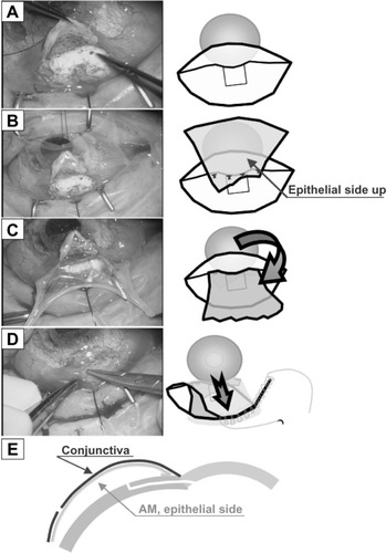
Table 1 Summary of the six cases of patients with refractory glaucoma and severe corneal disorders
Figure 2 Images of three representative cases.
Notes: (A–C) Filtering blebs post AM-assisted TLE in all three patients. (D) Optical coherence tomography image (sagittal view) of the filtering bleb in case 1 at 2 years postoperative.
Abbreviations: AM, amniotic membrane; TLE, trabeculectomy.
Abbreviations: AM, amniotic membrane; TLE, trabeculectomy.
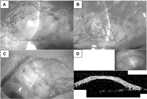
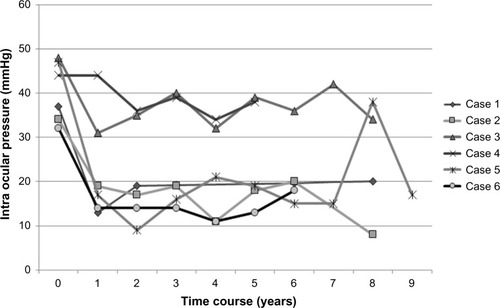
Figure 3 Time course of IOP changes in all six cases.
Abbreviation: IOP, intraocular pressure.