Figures & data
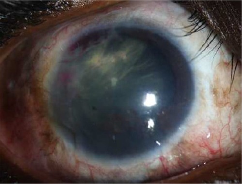
Figure 1 A well-circumscribed amelanotic iris mass with underlying multiple posterior pigment epithelial cysts.
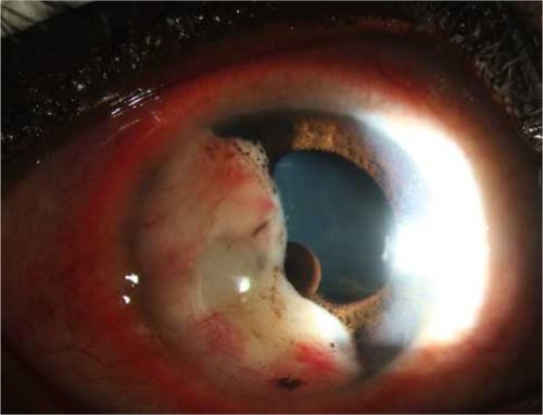
Figure 2 UBM revealed a hypoechoic mass coalesced with iris stroma with 5.6 mm horizontal diameter and 9.3 mm vertical diameter.
Abbreviation: UBM, ultrasound biomicroscopy.
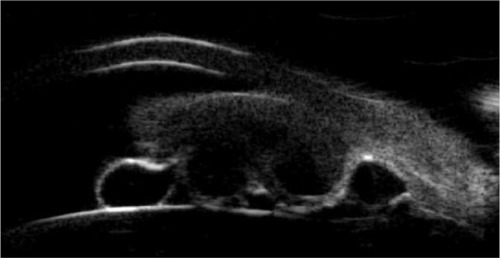
Figure 3 Histopathology and Immunohistochemical stain.
Notes: (A) Histopathology of the surgical specimen showing attached iridic tissue (H&E ×40). (B) Tumor cells having plump shape and epithelioid appearance with mildly pleomorphic and vesicular nuclei (H&E ×400). (C) Immunohistochemical stain for alpha smooth muscle actin (IHC ×100). (D) In situ hybridization for EBER shows positive reaction (EBER ×400).
Abbreviations: EBER, Epstein–Barr virus-encoded RNA; H&E, hematoxylin and eosin; IHC, immunohistochemistry.
Abbreviations: EBER, Epstein–Barr virus-encoded RNA; H&E, hematoxylin and eosin; IHC, immunohistochemistry.
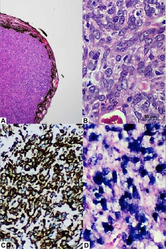
Figure 4 Final slit-lamp biomicroscopy at 8 months revealed the absence of tumor recurrence.