Figures & data
Figure 1 (A) shows acute cilioretinal artery occlusion after 24 hours following cardiac catheterization. (B) shows resolution of the retinal edema, and residual retinal exudates, 14 weeks later.
Notes: Note deep retinal edema extending into the fovea of the left eye. Present also is a choroidal nevus of the macula.
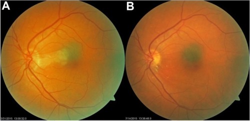
Figure 2 OCT scan of retina in cross-section showing loss of RPE and deep retinal structures nasal to the fovea 14 weeks after the occlusion.
Abbreviations: OCT, optical coherence tomography; RPE, retinal pigment epithelium.
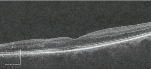
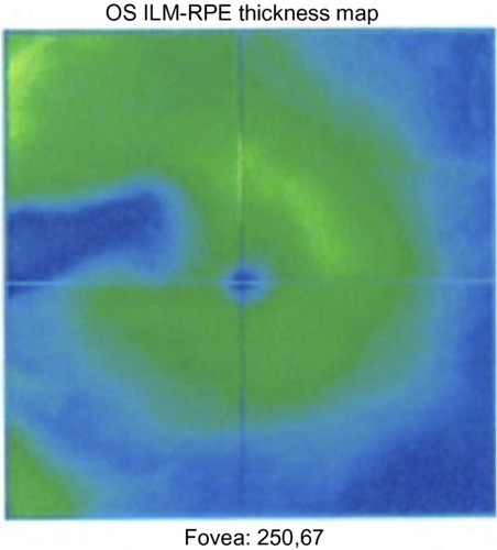
Figure 3 OCT scan of the macula showing loss of retinal pigment epithelium 14 weeks after the occlusion.
Abbreviations: OS, oculus sinister; ILM, internal limiting membrane; RPE, retinal pigment epithelium; OCT, optical coherence tomography.