Figures & data
Figure 1 Clinical photographs of the right eye.
Notes: Arrow in (A) points to a mass lesion below the eyebrow. Arrow in (B) points to a well-localized mass over the subcutaneous region. (C) The yellowish mass is 2 cm × 1 cm in size with irregular border and rubbery consistency.
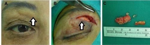
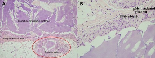
Figure 2 Microscopic examinations.
Notes: (A) Foreign body granuloma composed of multiple nodules characterized by basophilic amorphous materials surrounded by an irregular fibrous border (H&E, ×100). (B) The fibrous border composed of histiocytes, multinucleated giant cells, and fibroblasts (H&E, ×400).
Abbreviation: H&E, hematoxylin and eosin.
Abbreviation: H&E, hematoxylin and eosin.