Figures & data
Table 1 Demographics, background patient and surgeon characteristics, past medical history, and outcomes of patients included in the series
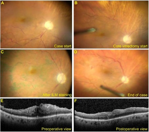
Figure 1 Intraoperative view of optic nerve and macular ischemia in Case 1 patient.
Notes: At the beginning of the case, prior to initiation of vitrectomy, marked optic nerve pallor, retinal vessel attenuation, and box-carring were noted (A). There was instantaneous but partial improvement in optic nerve and macular perfusion as soon as the vitreous cutter was engaged (B). The perfusion continued to improve throughout the procedure, as shown after ILM staining (C) and after membrane peeling (D). Macular OCT shows preoperative ERM (E). At 6-week follow-up, there is improved macular thickening and resolution of intraretinal cysts but significantly worsened visual acuity (F).
Abbreviations: ERM, epiretinal membrane; ILM, internal limiting membrane; OCT, optical coherence tomography.
Abbreviations: ERM, epiretinal membrane; ILM, internal limiting membrane; OCT, optical coherence tomography.