Figures & data
Table 1 A few cases with macular tear have been previously reported in the literature. Our current case is the first report of an idiopathic case
Figure 1 (A) Ophthalmoscopy revealed a horseshoe-like tear on the temporal side of the macula in the left eye. (B) Fluorescein angiography revealed a non-perfusion area and that there was no retinal vein occlusion. (C) Spectral-domain optical coherence tomography revealed the focal retinal detachment in the left eye reached the fovea.
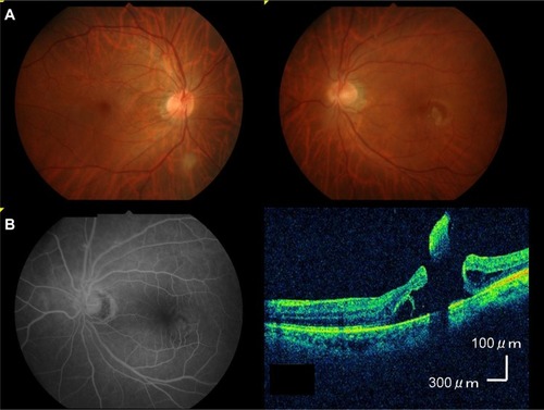
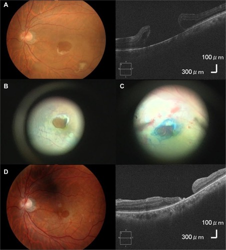
Figure 2 (A) A few days after the first visit, there was no longer adhesion of the flap of the tear to the retina and the tear size had increased to 1.5 disc diameters. (B) Before the surgery, the macular tear size was 1.5 disc diameters. (C) In order to close the tear, we performed the inverted internal limiting membrane flap technique with 20% SF6 gas tamponade. (D) Although the disc diameter of the tear after the surgery decreased to 0.5 disc diameters, complete closure of the tear was not achieved.