Figures & data
Figure 1 Ocular surface before and after necrotic tissue excision.
Notes: (A) Ocular surface 10 days after an acute burn. An extensive necrotic/degenerative corneoconjunctival tissue covering the entire surface is shown. (B) Under surgery microscope, a perfectly clear normal Descemet’s membrane was observed.
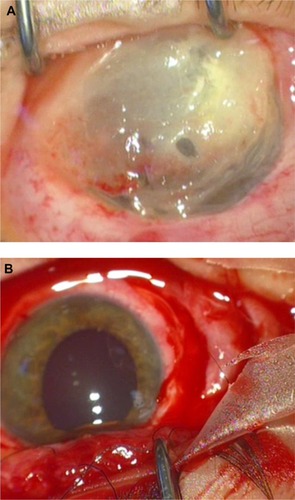
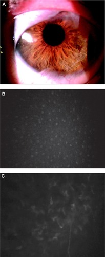
Figure 2 Postoperative evaluation by means of slit-lamp and corneal confocal microscopy.
Notes: (A) Postsurgery evaluation (4–5 months). On slit-lamp biomicroscopy, the cornea and anterior chamber were both clear with restored corneal stromal over the follow-up period. (B) Confocal microscopy shows epithelial and stromal regeneration with reinnervation. A normal corneal epithelium morphology with large superficial cells is seen in the regenerated epithelium. (C) Corneal nerve bundles regenerated in the corneal stroma. Anterior keratocytes and stroma were unevenly distributed, with bright nuclei and signs of opacity. (B and C) Corneal confocal microscopy was performed using Confoscan 4.0 (Nidek, Gamogori, Japan), equipped with an Achroplan non-applanating 40× immersion objective lens (Zeiss, Oberkochen, Germany). The standard dimension of each image produced was 420×315 µm (area=0.132 mm2), with an optical section thickness of 5 µm.